Medical diagnostic errors affect about 12 million Americans yearly. Misinterpreted diagnostic images add substantially to this concerning number. Patient outcomes, treatment decisions, and lives depend on medical imaging quality. Computer-based diagnostic systems have shown they can cut down interpretation errors by up to 30% for specific imaging tasks.
The medical imaging technology landscape keeps changing faster than ever. Experts predict the global AI-based medical image analysis market will hit $7.5 billion by 2034. The market shows a steady 7.9% compound annual growth rate. These numbers highlight quality image diagrams’ growing role in modern healthcare. Regional centers of excellence, like diag imagerie facilities in France – diag imagerie choisy le roi and diag imagerie bois colombes, help set global standards. Healthcare institutions that use advanced diagnostic image solutions have seen fewer diagnostic errors. They also interpret images 40% faster.
This piece will get into why image quality matters from a medical professional’s point of view. We’ll look at technical components, clinical implications, and new technologies that will change diagnostic imaging in 2025 and beyond.
Understanding the Workflow Behind Diag Image Creation
Creating diagnostic images involves many steps that affect image quality. Medical professionals must pay close attention to detail at each stage to get accurate diagnoses. A good grasp of this process helps doctors spot issues that could affect diagnostic accuracy.

Patient preparation and positioning
Quality diagnostic images need proper patient preparation. Each imaging type needs specific protocols to get the best images with minimal artifacts. To name just one example, CT scans need patients to fast for about 4 hours before the scan. MRI cardiac imaging requires patients to avoid caffeine 24 hours before the procedure.
Preparation instructions typically include:
- Dietary restrictions (fasting requirements)
- Medication adjustments
- Removal of metallic objects
- Specific positioning guidelines
- Use of contrast media when needed
Technologists play a significant role. They check patient identity, explain procedures, and make sure positioning is correct. The right position makes a big difference in image quality. Small positioning errors can make images useless for diagnosis. Different body parts need standard views (anteroposterior, lateral, and oblique) with specific techniques.
Image acquisition process
A radiologic technologist captures the diagnostic information after positioning the patient. They use advanced equipment during this phase. The process varies by a lot between X-ray, CT, MRI, and ultrasound.
Technical settings during acquisition affect image quality. X-ray technologists adjust kVp (kilovoltage peak) and mAs (milliampere-seconds) to control contrast and optical density. CT scan parameters like slice thickness control spatial resolution in the longitudinal direction. This creates important tradeoffs between resolution, noise, and radiation dose.
Source-image distance (SID) affects image contrast, distortion, and magnification. The imaging process must follow standard protocols while meeting individual patient needs. This balance gives consistent quality for unique clinical cases.
Data reconstruction and post-processing
Image reconstruction turns physical data from imaging equipment into diagnostic images. These images show tissue properties in the spatial domain. This process works as the inverse of mapping tissue to acquired data through physics models.
Each modality uses different reconstruction methods:
- MRI uses inverse fast Fourier transform (IFFT)
- CT applies filtered back projection (FBP)
- PET uses iterative reconstruction (IR)
Radiologists often need faster scans to improve patient safety and throughput. This means collecting incomplete data, which makes reconstruction harder. They need prior knowledge to reduce noise and artifacts from these quick scans.
Deep-learning (DL)-based reconstruction methods have shown great results in reducing noise and artifacts. But these methods can struggle with different organ sizes and appearances. This might lead to over-smoothing and loss of anatomical details. Hybrid DL-IR (deep learning-iterative reconstruction) schemes now use the best parts of both methods.
Post-processing is the final step. Images get enhanced to help with diagnosis. Common post-processing includes windowing, spatial filtering, edge enhancement, and noise reduction. These changes make images better for radiologists to diagnose conditions.
Both acquisition methods and reconstruction algorithms affect the quality of final images. This quality directly affects how accurate and consistent diagnoses will be.
Technical Components That Define Image Quality
Three key technical factors determine what clinicians can—and cannot—detect in diagnostic images. Every part of the imaging chain, from radiation-capturing physical components to data-processing software, plays a vital role in diagnostic accuracy.
Sensor resolution and detector type
The detector forms the basis of any diagnostic image. It captures incoming radiation as a physical component. Modern imaging systems use two main detector types: direct and indirect conversion systems. Direct conversion detectors use photoconductors like amorphous selenium (a-Se) to convert X-rays into electrical signals. Indirect systems work differently. They first change radiation to visible light through scintillators, then transform this light into electrical signals using photodiodes.
Detector elements’ physical size directly affects spatial resolution. Digital radiography typically uses matrix sizes of 2000 × 2500, with pixel sizes around 175 μm for a 35 × 43-cm cassette. Digital mammography needs even higher resolution, using pixel sizes between 50 and 100 μm and matrix sizes of approximately 3000 × 4000. A mammogram contains about 12 million pixels, which requires special display technologies or zoom features to see all details.
Photon-counting detector CT (PCD-CT) has emerged as an advanced technology that solves many problems of conventional energy-integrated detectors. PCD-CT uses semiconductor materials to generate signals matching each photon’s energy, which allows better energy discrimination. This technology brings several benefits:
- Better spatial resolution through smaller detector pixel sizes
- Improved signal-to-noise ratios by eliminating electronic noise
- More effective detection of low-energy photons needed for image detail
Software algorithms and image compression
Software algorithms transform raw data into useful diagnostic images. Reconstruction algorithms create the final image from the original signals. Traditional filtered back projection (FBP) methods have given way to iterative reconstruction (IR) techniques and deep learning image reconstruction (DLIR) methods.
DLIR uses deep neural networks trained on high-quality images to separate noise from signals. This suppresses unwanted artifacts while preserving anatomical structures. Research shows that DLIR can improve image quality even with less radiation and contrast medium—a big step forward for patient safety.
Image compression is another key part of the technical setup. Medical images take up lots of storage space, so efficient compression helps with both archiving and transmission. The DICOM standard supports various compression formats:
- JPEG (baseline and extended)
- JPEG-LS for near-lossless compression
- JPEG2000 offering resolution and quality scalability
- MPEG formats for video sequences
Medical applications prefer lossless compression because it keeps all original pixel data intact, though compression ratios usually stay between 2:1 and 4:1. Soft compression methods can achieve higher ratios and have shown better results than traditional approaches.
DICOM standards and metadata integrity
The Digital Imaging and Communications in Medicine (DICOM) standard is the life-blood of medical image management. DICOM does more than store pixel data—it includes essential metadata that provides context for accurate diagnosis. Images stay connected to their descriptive information, following the standard’s rule that “the image can never be separated from this information by mistake”.
DICOM metadata contains important acquisition parameters for each modality. CT images include X-ray tube voltage, current, exposure time, and slice thickness information. Nuclear medicine studies record details about radiopharmaceuticals, injection times, and how long the acquisition took. Doctors need this context to interpret images correctly.
DICOM metadata uses a standardized format with attributes identified by unique tags. Each attribute has a group number and element number in hexadecimal format, like (0008, 0060) for modality type. This standardization helps different vendors and facilities work together, which benefits diagnostic imaging centers in France and worldwide.
Metadata quality directly affects AI and machine learning in radiology. Incomplete or poor metadata can create bias in algorithm training, which might lead to dangerous misinterpretations. That’s why facilities like diag imagerie bois colombes must maintain strict metadata standards for both human diagnosis and new AI applications.
Clinical Implications of High vs. Low Image Quality
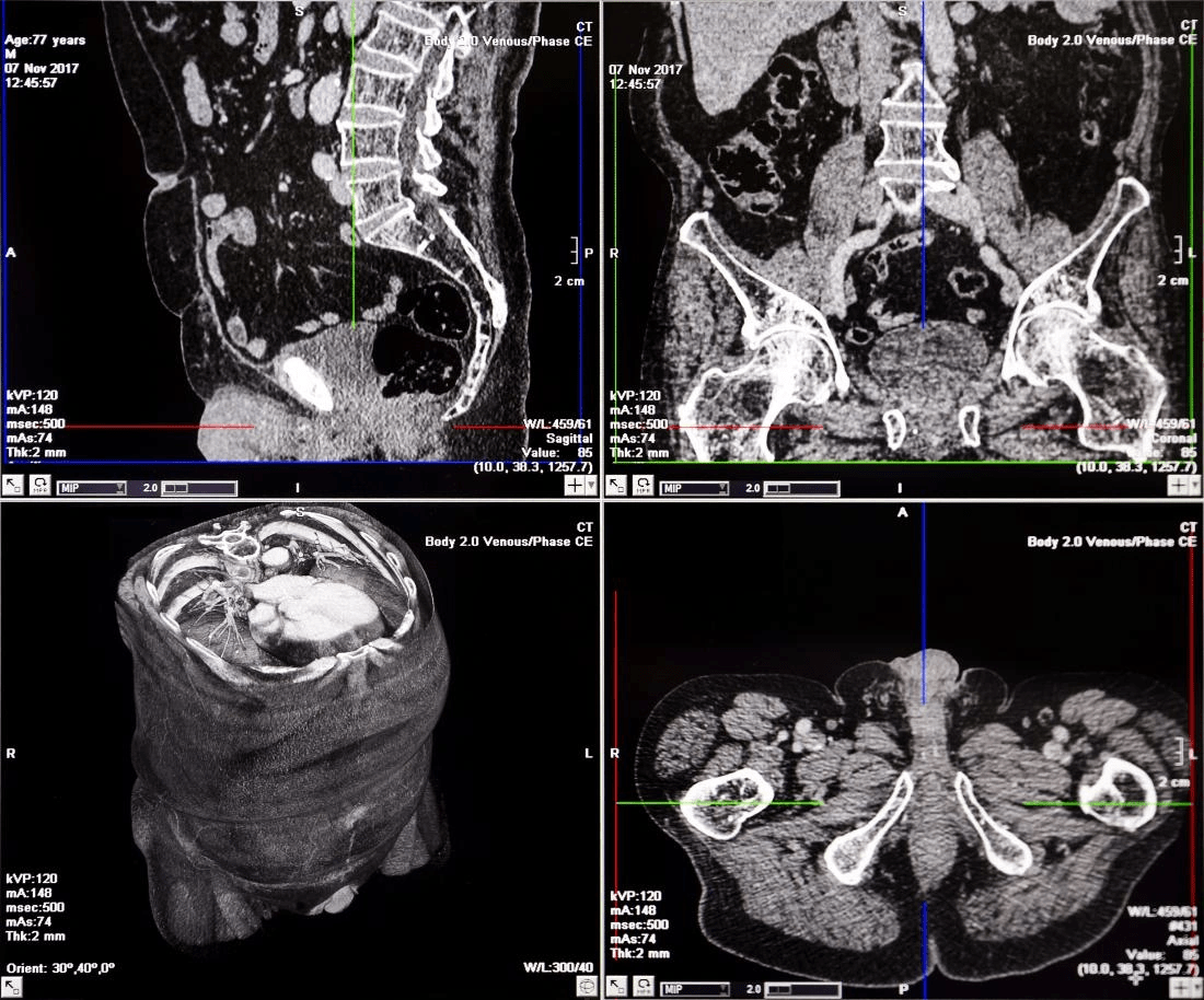
Image Source: Medical News Today
The quality of diagnostic images can mean the difference between life and death in clinical settings. Poor image quality makes accurate interpretation harder, which leads to wrong diagnoses, takes longer to interpret, and ends up putting patient care at risk.
Case study: Stroke misdiagnosis due to poor CT quality
Image quality plays a vital role in patient outcomes, especially for stroke diagnosis. Quick treatment is essential for acute ischemic stroke patients, and accurate brain imaging helps doctors manage these cases properly. CT scans are common but often fail to detect strokes reliably—especially during the first 48 hours when ischemic changes look normal.
Research shows this dangerous problem clearly. 24 out of 32 patients showed negative CT results at first, which led to wrong diagnoses. In fact, when doctors compared different scanning methods, CT only found cerebellar infarction in 45.2% of cases. MRI proved much better, detecting all cases no matter when doctors took the scan after symptoms started.
This diagnostic challenge exists because noncontrast CT works well to rule out brain bleeding but doesn’t catch early signs of brain ischemia in the first 6 hours. Even advanced CT protocols aren’t very sensitive (25.6%) to stroke mimics, though they rarely give false positives.
Impact on surgical planning and treatment decisions
Image quality affects surgical planning and treatment in every medical specialty. Low-quality images create several problems that get worse over time:
- Extended reporting times: Unclear images force clinicians to adjust settings, look at old scans, or check more sequences, which slows down diagnosis
- Decision delays: Doctors often wait for better images before making recommendations, which affects acute care patients the most
- Increased resource utilization: Unclear scans mean more follow-up tests and extra work for staff in already busy hospitals
Image resolution remains a basic challenge. Even with advanced MRI and CT machines, some conditions are hard to see clearly. Small tumors in early stages might go unnoticed, which leads to incomplete surgical plans. Metal implants from earlier surgeries can also create image artifacts that make interpretation difficult.
Research proves the link between quality and diagnostic confidence. A study of skin condition diagnoses using mobile phone photos showed that poor image quality dropped accuracy to 50% compared to in-person exams.
Legal and ethical considerations
Legal issues around image quality raise concerns about patient privacy, consent, and possible lawsuits. About 75% of malpractice lawsuits against radiologists involve diagnostic errors.
Patient consent becomes crucial for clinical images. Medical teams need to discuss several points with patients:
- The exact purpose of taking clinical images
- Ways the images might be used (including training or research)
- People who will see the images
- Methods to keep images anonymous
Bad photo technique can make surgical outcomes look wrong and might not count as evidence in court. Doctors and healthcare facilities must protect patient confidentiality, including diagnostic images. Medical teams need to keep watching and adapting to social and scientific changes to maintain ethical standards in diagnostic imaging.
AI and Machine Learning in Image Quality Control
AI has changed quality control in medical imaging faster than ever. The FDA has authorized over 500 AI-enabled devices for clinical use, including 91 in 2022 alone. This growth shows how AI helps improve diagnostic image quality in medical facilities worldwide, including specialized centers like diag imagerie bois colombes.
Real-time quality assessment tools
Modern diagnostic imaging uses deep learning algorithms that check image quality as they’re taken and flag issues immediately. These systems get better as they process more data. They become fluent in spotting differences between normal variations and concerning anomalies.
These tools make a big difference. AI-enabled X-ray image processing gives sharp detail and stays consistent even when exposure techniques vary or exam conditions are tough. This simplified process means fewer repeat exams, lower rejection rates, and fewer patient positioning errors.
Deep Learning Image Reconstruction (DLIR) brings another breakthrough to immediate quality improvement. Recent studies show DLIR improves image quality more than traditional reconstruction methods:
- Up to 54% increase in signal-to-noise ratio for gray matter
- 60% increase for white matter
- 58% enhancement in contrast-to-noise ratio in basal ganglia structures
AI-based quality control goes beyond radiography. Deep learning approaches to PET image noise reduction show better lesion outlines and overall image quality compared to standard reconstruction.
AI in diag image france and global adoption
French diag image centers welcome AI-powered quality control systems more and more, following global trends. French facilities like diag imagerie choisy le roi add these technologies to their daily work.
AI applications in diagnostic imaging keep growing. The FDA has doubled their review of AI and machine learning-enabled devices from 2017 to 2018, and growth continues. The FDA authorized 115 submissions in 2021, which is 83% more than 2018. This growth shows increasing trust in AI’s power to improve image quality and diagnostic accuracy.
A recent meta-analysis of five clinical validation studies found AI-based interventions improved image quality by a lot (mean difference 0.70, 95% CI 0.43-0.96; P<.001). CT technology advances show promising results to reduce radiation dose, though these findings weren’t statistically significant (mean difference 0.47, 95% CI -0.21 to 1.15; P=.18).
Bias and training data challenges
Despite progress, AI systems face big challenges with bias and training data. Developers and users must spot potential biases throughout the AI system development pipeline during training, validation, and testing.
Several critical bias sources need attention. Small training datasets that don’t show the variety of real-life cases create a basic challenge. Large datasets can also show bias if they don’t properly include:
- Patient demographics
- Imaging equipment variations
- Institution-specific protocols
- Temporal factors affecting disease presentation
Experts call these issues “domain shift” – where AI systems don’t work as well because development data doesn’t match deployment environments. In stark comparison to this, data leakage happens when training images accidentally appear in test sets, making performance look better than it is.
Technical and ethical concerns both need attention. A systematic review found none of 62 AI models developed to detect COVID-19 using X-ray and CT images could work in clinical practice due to method flaws. Quality control must extend to AI systems themselves to ensure they improve rather than hurt diagnostic accuracy.
Quality Assurance in Diag Imagerie Centers
Quality assurance is the life-blood of diagnostic imaging excellence. Healthcare facilities need systematic procedures to maintain consistent performance standards. A well-designed QA program ensures patient safety and diagnostic accuracy by monitoring and evaluating all imaging processes.
Role of diag imagerie bois colombes and similar centers
Specialized facilities like diag imagerie bois colombes set the standard in implementing detailed quality assurance frameworks. These centers actively pursue the Quality Standard for Imaging (QSI)—a distinctive mark of excellence that The Royal College of Radiologists and The College of Radiographers developed. QSI gives providers a well-laid-out approach to evaluate performance and improve patient care outcomes.
Diagnostic imaging centers of all sizes throughout France work to establish resilient quality management systems. Quality committees supervise these systems with a team of radiologists, radiographers, medical physicists, sonographers, and administrative staff. The committees focus on:
- Compliance with regulatory standards
- Equipment performance optimization
- Patient-focused care delivery
- Building teamwork in all departments
Routine audits and compliance checks
Clinical audit is a mandatory legal requirement in the European Union based on the European Commission Basic Safety Standards Directive. The Royal College of Radiologists developed AuditLive, which offers many audit templates that departments can adapt locally.
These audits follow a cycle: planning resources, selecting good practice standards, measuring performance, making changes, and re-auditing to check improvements. Audit topics cover everything from image quality assessment to protocol adherence and radiation dose monitoring.
The European Society of Radiology’s expanded AUDITRAD system now includes 33 new audit templates and 55 revised ones. These templates cover significant contemporary topics including AI tool usage. This standardized approach helps imaging departments maintain consistent quality in their practices.
Staff training and certification
Education is vital to quality assurance in diagnostic imaging. Through collaboration with Health Education England, the College of Radiographers developed Clinical Imaging e-learning. This platform supports radiographers at different career stages. Staff members can maintain their imaging skills in radiography, CT, MRI, and ultrasound while completing professional development activities.
The Midlands Imaging Academy and similar training academies expand regional capacity through multi-professional training. Electronic picture archiving suites are a great way to get remote training and enable collaborative efforts between education centers.
Certification proves competency and adherence to professional standards for radiologic technologists. Professionals must complete 24 hours of continuing education every two years to keep their certification current.
Future Trends in Diag Image Quality Enhancement
Image Source: Rayscape
Medical imaging has entered a new era where advanced technologies redefine how doctors capture, process, and use diagnostic images.
3D imaging and augmented reality
AR technology changes diagnostics by adding digital data to the real world. Healthcare’s global market for AR and VR reached USD 1.57 billion in 2022 and will grow to USD 13.74 billion by 2032, with a 24.81% annual growth rate. AR-assisted image-guided biopsies perform better than traditional techniques, particularly with smaller targets. Doctors can now create and manipulate 3D renderings from 2D scans. They rotate, zoom, and cross-section images using VR headsets. To name just one example, EchoPixel True 3D allows detailed examination through standard VR devices.
Edge computing and 5G-enabled diagnostics
Fifth-generation mobile technology has altered the map of diagnostic image transmission with these capabilities:
- Data speeds up to 10 Gbps (100 times faster than 4G)
- Very low latency (1ms)
- 1000 times higher capacity
5G helps transmit mobile ultrasound from ambulances to hospitals with average throughputs of 4 Mbps and round-trip latencies of just 10 milliseconds in real-life applications. High-performance edge computing combined with 5G allows immediate processing of data-intensive imaging workloads. The edge computing healthcare market reached USD 5.28 billion in 2023.
Personalized imaging protocols
Customized imaging marks a radical alteration from “one-protocol-fits-all” practices to patient-specific examinations. Doctors should optimize imaging protocols based on lesion site, anatomy, gender, age, imaging history, and clinical requirements. Patient-specific approaches use site-specific and size-specific protocols while maintaining diagnostic quality, not just reducing radiation exposure. Mobile applications and big data tools in clinical workflows help doctors make better decisions.
Conclusion
Image quality is the life-blood of modern healthcare delivery. Our analysis shows how each part of the imaging process affects diagnostic accuracy and patient outcomes. This includes everything from patient positioning to data reconstruction. Healthcare providers need to understand that even small technical issues can affect clinical decisions and treatment success.
Imaging technology continues to advance rapidly. New developments in sensor technology, reconstruction algorithms, and AI-powered quality control tools have improved diagnostic capabilities. These advances have also reduced radiation exposure and interpretation time. Centers like diag imagerie show how dedication to quality standards lifts excellence in medical imaging.
Poor image quality can have serious clinical effects. Doctors might miss crucial diagnoses, especially when dealing with time-sensitive conditions like stroke. This can lead to devastating risks for patients. The issue goes beyond clinical concerns into legal and ethical areas. Healthcare providers could face lawsuits while raising questions about patient consent and privacy.
AI has become a game-changer in diagnostic imaging quality. New tools can spot issues during image capture. Deep learning methods now create clearer images than traditional approaches. These technologies show promise but face challenges with bias and limited training data. We need to solve these issues before widespread adoption.
The combination of 3D imaging, augmented reality, and 5G will change diagnostic capabilities even more. Standardized approaches will give way to personalized imaging protocols. These will offer custom examinations based on patient characteristics without losing diagnostic accuracy.
Healthcare providers must treat diagnostic imaging quality as both a technical and ethical priority. Quality assurance programs, regular audits, and ongoing professional education are vital investments, not optional extras. Excellence in medical imaging quality serves healthcare’s highest purpose – better patient outcomes through accurate and timely diagnosis.
FAQs
1. Why is image quality so important in medical diagnostics?
Image quality is crucial in medical diagnostics because it directly impacts the accuracy of diagnoses, treatment decisions, and patient outcomes. High-quality images allow medical professionals to detect subtle abnormalities, reduce interpretation errors, and make more informed clinical decisions.
2. How does AI contribute to improving diagnostic image quality?
AI enhances diagnostic image quality through real-time quality assessment tools, deep learning reconstruction methods, and automated artifact reduction. These technologies can improve image clarity, reduce noise, and help identify potential issues during image acquisition, ultimately leading to more accurate diagnoses.
3. What are the clinical implications of poor image quality?
Poor image quality can lead to missed or delayed diagnoses, unnecessary follow-up examinations, and incorrect treatment decisions. In time-sensitive conditions like stroke, suboptimal image quality can have severe consequences for patient outcomes and may even result in legal issues for healthcare providers.
4. How are personalized imaging protocols improving patient care?
Personalized imaging protocols tailor examinations to individual patient characteristics such as age, gender, anatomy, and clinical requirements. This approach optimizes image quality while minimizing radiation exposure, resulting in more accurate diagnoses and improved patient safety.
5. What role do quality assurance programs play in diagnostic imaging centers?
Quality assurance programs in diagnostic imaging centers ensure consistent performance standards, patient safety, and diagnostic accuracy. These programs involve regular audits, compliance checks, staff training, and certification to maintain high-quality imaging services and improve patient care outcomes.